搜索更多信息
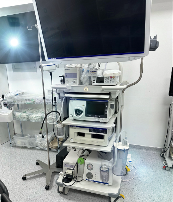
视频处理器 奥林巴斯 CV-1500。 视频胃镜 奥林巴斯 GIF-EZ1500。 奥林巴斯 CF-EZ1500DL。 视频监视器 奥林巴斯 OEV321UH。 机架式内窥镜 奥林巴斯 WM-NP3。
370 x 198 x 488 mm 398 x 218 x 580 mm (maximum) 19.4 kg
4K 超高清成像 窄带成像 (NBI) 反应灵敏的触摸屏界面 多功能内窥镜兼容性 先进的图像处理 耐用可靠的设计
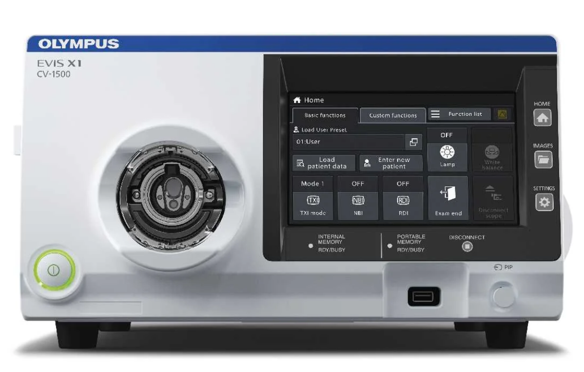

Olympus EVIS X1 CV-1500 Video System Center Experience Precision and Clarity with the Olympus Evis X1. Explore 4K Ultra-High-Definition Imaging, NBI, and Workflow Efficiency for Enhanced Endoscopic Procedures.
奥林巴斯 EVIS X1 CV-1500 视频系统中心 使用奥林巴斯 Evis X1 体验精确和清晰。探索 4K 超高清成像、NBI 和工作流程效率,增强内窥镜手术。

说明
EVIS X1 是一款开创性的医疗成像系统,为世界内窥镜检查设立了新标准。这款先进的设备采用尖端技术,为医疗专业人员提供了在诊断和治疗过程中卓越的视觉清晰度和精确度。
“Evis X1 非常直观,为内窥镜医生提供了一系列创新的、经过验证的工具,使他们能够进行微创、精确和有效的手术,从而做出真正明智的诊断和治疗决定,为患者带来最大利益。在这些新技术中,TXI 看起来尤其有前途。它似乎能为内镜医师提供更多信息,同时保持接近白光的外观。这将使我们很容易适应它"。

Michal Kaminski, National Research Institute of Oncology in Warsaw
EVIS X1 专为早期抗癌而设计,可对癌前病变进行可靠、准确的诊断,从而实现精确、简便的筛查。在内窥镜系统中,EVIS X1 引入了最新的、易于使用的技术来识别、评估和治疗胃肠道疾病,如结直肠癌(CRC)和呼吸系统疾病。作为技术市场的领导者,奥林巴斯不断开发包括人工智能(AI)在内的未来技术。
EVIS X1 主要功能:
4K 超高清成像: Evis X1提供4K超高清成像,提供无与伦比的视觉细节和实时清晰度。这使医疗从业人员能够非常精确地观察组织和结构,提高诊断的准确性。
窄带成像(NBI): 窄带成像技术提高了血管和粘膜形态的可视化程度,使检测细微病变和异常变得更加容易。这一功能对于早期诊断和有效治疗至关重要。
反应灵敏的触摸屏界面: 该系统拥有直观、反应灵敏的触摸屏界面,确保医疗专业人员易于使用。它简化了操作,使医生能够专注于病人护理。
多功能内窥镜兼容性: Evis X1 兼容多种奥林巴斯内窥镜,可适应不同医疗专业的各种手术要求。
先进的图像处理: 该系统采用了优化图像的算法。

优点
卓越的视觉清晰度: Evis X1 采用 4K UHD 成像和 NBI 技术,为医疗专业人员提供无与伦比的视觉清晰度,有助于精确诊断和有效治疗。
高效的工作流程: 反应灵敏的触摸屏界面和用户友好型控件简化了内窥镜检查流程,减少了患者的不适感,缩短了手术时间。
经济高效: 该系统的耐用性和可靠性有助于降低维护和更换成本,使其成为医疗机构的高性价比之选。
适应性强: Evis X1 与各种内窥镜的兼容性确保其适用于各种医疗程序,使其成为医疗机构的多功能解决方案。
临床信心: 医疗保健专业人员可以对 Evis X1 充满信心,因为他们知道自己可以使用先进的技术,获得卓越的成像质量。
EVIS X1 通过引入易于使用的新技术,帮助胃肠道疾病(如结肠直肠癌 (CRC))或支气管疾病的检测、定性和治疗,从而支持准确的筛查和可靠的诊断。
这些技术包括扩展景深成像(EDOF)、红色双色成像(RDI)、纹理和色彩增强成像(TXI),以及已经广为人知并得到验证的窄带成像(NBI)。奥林巴斯致力于创新,也在开发以人工智能(AI)为主要特征的未来技术。

Include:
Video processor Olympus CV-1500.
Videogastroscope Olympus GIF-EZ1500.
Videocolonoscope Olympus CF-EZ1500DL.
video monitor Olympus OEV321UH.
Rack endoscopic Olympus WM-NP3.

Specifications:
| Power Supply | Rated voltage | 100-240 V AC; Within ±10% |
| Frequency | 50/60 Hz; within ±3 Hz | |
| Rated input | 600 VA | |
| Size | Dimension (W x H x D) | 370 x 198 x 488 mm |
| 398 x 218 x 580 mm (maximum) | ||
| Weight | 19.4 kg | |
| Classification (Medical Electrical Equipment) | Type of protection against electric shock | Class I |
| Degree of protection against electric shock of applied part | Depend on applied part. (The degree of protection against electric shock of this product is BF type if the mounting part to be connected to this product is BF type. However CF type is not subject to combination in this product.) | |
| Degree or protection against explosion | The video system center should be kept away from flammable gases. | |
| Observation | Analog signal output | VBS composite |
| Digital signal output | 12G-SDI 3G-SDI HD-SDI SD-SDI | |
| User settings | The function settings for up to 20 users can be stored. | |
| Color tone adjustment | Adjust the color tone of each endoscopic image for White light observation mode, NBI Technology observation mode, and RDI Technology observation mode Red adjustment : ±8 steps · Blue adjustment : ±8 steps · Chroma adjustment : ±8 steps. | |
| Automatic gain control (AGC) | The image can be electronically amplified when the light is inadequate due to the distal end of the endoscope being too far from the object | |
| Contrast | H (High): Darkens the dark part and brightens the bright part. L (Low): Brightens the dark part and darkens the bright part. | |
| BAI-MAC | Brightness adjustment with the maintenance of contrast | |
| Iris | The iris modes can be switched. · Auto: The brightness is adjusted based on the central part’s brightest part and the periphery part’s average brightness. · Peak: The brightness is adjusted based on the brightest part of the endoscopic image. Average: The brightness is adjusted based on the average brightness of the endoscopic image. | |
| Image enhancement settings | Fine patterns or edges in the endoscopic images can be enhanced electrically to increase the image’s sharpness. 1、 Enhancement type A : Emphasizes the pattern and contour of the endoscopic image. 2、Enhancement type B : Emphasizes the finer parts than structure emphasis type A. | |
| Switching the enhancement modes | The enhancement level can be selected from 3 levels (OFF, 1, 2, and 3) | |
| Image size selection | The size of the endoscopic image can be selected from 2 modes. (Except SDTV) |
| Electric zoom | Switch between mode 1, mode 2, and mode 3. | |
| PIP/POP | Switch between PIP and POP. | |
| Aspect ratio | Switch between 16:9 and 4:3. (Except SDTV) | |
| Freeze | Freeze the endoscopic image. | |
| Pre-freeze | The image with the least blur is selected from the images captured in the set time period before freeze operation and displayed. | |
| Optical-digital observation | The optical-digital observation can be performed. An endoscope compatible with the optical-digital observation is required. NBI Technology observation: This observation mode uses the narrow-band light. · RDI Technology observation: This observation mode uses the red dichromatic lights. · TXI Technology observation: This observation mode enhances color, texture and brightness. | |
| Beginning and ending examination | Beginning and ending examination timing can be set interlock with the particular operation. | |
| Custom switch | Assign specific functions to the following buttons. · Remote switches (Up to 5) · Foot switches (Up to 2) · Keyboard custom key (Up to 4) · Touch panel custom button of basic functions screen (Up to 3) · Touch panel custom button of custom functions screen (Up to 10) | |
| MyCV mode | Switch setting values of multiple functions at once. | |
| Documentation | Remote control | The following peripheral device can be controlled (specified models only). 1、Portable memory · Video recorder · Color video printer · Image filing system · Server |
| Patient information | The following data can be displayed on the monitor. · Patient ID · Patient name · Gender · Age · Date of birth · Comment | |
| Displaying the record state | The recording state of the following peripheral device can be displayed on the monitor. · Portable memory: Remaining capacity 1、Video recorder: Number of shots / Recording status · Color video printer: Number of shots · Image filing system: Number of shots | |
| Displaying the image information | The following data can be displayed on the monitor. · Image enhancement · Electric zoom ratio · Color mode · Focus · Observation mode | |
| Advanced registration of patient information | Up to 50 patient information can be registered. · Patient ID · Patient name · Gender · Age · Date of birth | |
| Recording format | Standard image quality: TIFF; Low image quality: JPEG | |
| Memory Backup | Memorization of user settings | The settings are held in memory even after the video system centre is turned OFF. |
| White balance | The white balance that is once set is held in memory (only when using the compatible endoscope) |
Conclusion:
The Olympus Evis X1 medical imaging system represents a significant leap forward in endoscopy technology. Its 4K UHD imaging, NBI capabilities, user-friendly design, and adaptability empower healthcare professionals to deliver exceptional patient care. Investing in the Evis X1 means investing in the future of healthcare, where precision and innovation lead to better patient outcomes.www.dzjxson.com